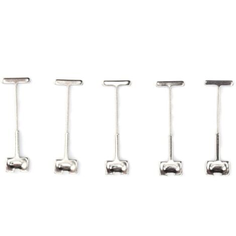
Left): Porcine ventricle sample, epicardium side up, mounted to
By A Mystery Man Writer
Description
Download scientific diagram | (Left): Porcine ventricle sample, epicardium side up, mounted to the silicone lined fixture with Tpins. (Right): Porcine aorta sample, intima side up, mounted to the silicone lined fixture with T-pins. (Both): 0.25 in diameter steel ball upper member as test probe. from publication: PolyJet 3D Printing of Tissue Mimicking Materials: An Investigation of Characteristic Properties of 3D Printed Synthetic Tissue | Current anatomical 3D printing has been primarily used for education, training, and surgical planning purposes. This is largely due to the models being printed in materials which excel at replicating macro-level organic geometries; however, these materials have the drawback | 3D Printing, Tissue and Subcutaneous Tissue | ResearchGate, the professional network for scientists.

Quantifying the microstructural and biomechanical changes in the
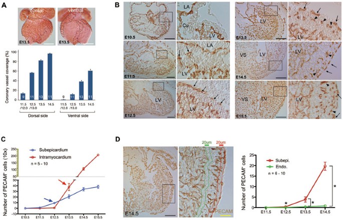
Subepicardial endothelial cells invade the embryonic ventricle
Frontiers Tipping the scales of understanding: An engineering

Heart Anatomy Anatomy and Physiology II

Medicina, Free Full-Text
Frontiers Preparing Excitable Cardiac Papillary Muscle and
Epicardial slices: an innovative 3D organotypic model to study

A Minimally Invasive, Translational Method to Deliver Hydrogels to
Biomimetics, Free Full-Text

Neuroanatomy of the Pig Cardiac Ventricles. A Stereomicroscopic
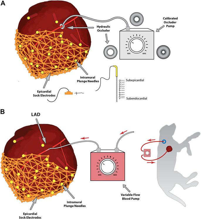
Frontiers Vagally-mediated heart block after myocardial

PDF) PolyJet 3D Printing of Tissue Mimicking Materials: An

Intramural Needle Ablation for Refractory Premature Ventricular

Neuroanatomy of the Pig Cardiac Ventricles. A Stereomicroscopic
Bioengineering, Free Full-Text
from
per adult (price varies by group size)